
Fluids and electrolytes are the water and dissolved ions that fill every compartment of the body, set osmotic gradients, and generate the electrical charges that drive nerve impulses and cardiac contractions. The single most important clinical rule: a sodium shift of just 10–15 mEq/L or a potassium change of 1–2 mEq/L can trigger seizures, coma, or a fatal arrhythmia. That is not hyperbole. It is why electrolyte panels are ordered on nearly every admitted patient, and why nurses are often the first to catch the subtle signs that precede a code.
Normal serum reference ranges to memorize (Chapter 15, Nursing Fundamentals):
- Sodium (Na+): 136–145 mEq/L — values below 120 mEq/L risk seizures and herniation
- Potassium (K+): 3.5–5.1 mEq/L — values above 6.5 mEq/L or below 2.5 mEq/L demand immediate ECG
- Calcium (Ca2+): 8.5–10.5 mg/dL — ionized calcium drives neuromuscular excitability
- Magnesium (Mg2+): 1.7–2.2 mg/dL — hypomagnesemia blocks potassium repletion and worsens arrhythmia risk
Always verify these against your facility's lab reference ranges, since analyzer methods vary.
Key Takeaways
Fluid and electrolyte balance is the foundation of safe nursing practice: small shifts in sodium or potassium cause life-threatening neurologic and cardiac events, and the correction rate matters as much as the correction itself.
Table of Contents
- What are fluid and electrolyte compartments?
- Major electrolytes: normal ranges, functions, and clinical pearls
- How does the body maintain fluid and electrolyte balance?
- Common electrolyte disorders: causes, symptoms, and red flags
- How to interpret labs and diagnose electrolyte disorders
- Treatment principles and management algorithms
- Nursing assessment, bedside calculations, and exam tips
- Complications and when to escalate
- Prevention and patient education
- Quick-reference tables and clinical algorithms
- What actually makes fluids and electrolytes click for nursing students
- Mastering fluids and electrolytes with Nursingschoolpro
- Sources
What are fluid and electrolyte compartments?
The body is mostly water. StatPearls puts total body water (TBW) at roughly 75% of body mass in infants and 50–60% in adults, with the difference explained by the lower water content of adipose tissue. Women and older adults tend toward the lower end of that range.
That water is not distributed evenly:
- Intracellular fluid (ICF): ~40% of body weight — the dominant compartment, inside every cell
- Extracellular fluid (ECF): ~20% of body weight, split into:
- Plasma represents a portion of extracellular fluid
- Interstitial fluid is a major part of extracellular fluid
- Transcellular fluid (CSF, synovial, pleural): the remaining small fraction
Osmolarity and why water moves
Plasma osmolarity typically falls within a normal physiologic range under stable conditions. Osmolarity is simply the concentration of solutes per liter of fluid. When that concentration rises on one side of a semipermeable membrane, water moves toward it by osmosis. Sodium is the dominant extracellular solute, so sodium concentration is the primary driver of plasma osmolarity and, by extension, of water distribution between compartments.
A quick clinical translation: give a patient a hypotonic IV fluid and you lower plasma osmolarity, pushing water into cells. Give a hypertonic saline and the reverse happens, pulling water out of cells into the vascular space. That physics is what makes fluid selection matter.
Starling forces at the capillary
Fluid also moves between plasma and the interstitium based on two opposing pressures. Hydrostatic pressure (the force of blood pushing outward) drives fluid out of capillaries. Oncotic pressure (the pull of plasma proteins, mainly albumin) draws fluid back in. When albumin drops, as in liver failure or malnutrition, oncotic pressure falls and fluid leaks into the interstitium, producing the edema you can pit with your thumb. Understanding this explains why giving normal saline to a hypoalbuminemic patient can worsen edema rather than fix it.
Major electrolytes: normal ranges, functions, and clinical pearls
Each electrolyte has a specific home, a specific job, and a specific way it signals trouble when it goes wrong. The table below gives you the numbers and the one-liner you need for clinical shifts and exams.
| Electrolyte | Normal Range | Primary Role | High-Yield Clinical Pearl |
|---|---|---|---|
| Sodium (Na+) | 136–145 mEq/L | Sets ECF osmolarity; drives water distribution | Hyponatremia is the most common inpatient electrolyte disorder; always check serum osmolality first |
| Potassium (K+) | 3.5–5.1 mEq/L | Resting membrane potential; cardiac repolarization | Hypokalemia flattens T waves and produces U waves; hyperkalemia peaks T waves and widens QRS |
| Calcium (Ca2+) | 8.5–10.5 mg/dL | Muscle contraction, coagulation, nerve excitability | Hypocalcemia causes Chvostek's and Trousseau's signs; hypercalcemia causes "bones, groans, moans, stones" |
| Magnesium (Mg2+) | 1.7–2.2 mg/dL | Enzyme cofactor; stabilizes cardiac membranes | Low Mg2+ blocks Na+/K+ ATPase, making hypokalemia refractory to potassium replacement alone |
| Chloride (Cl-) | 136–145 mEq/L | Acid-base balance; pairs with Na+ for osmolarity | Hyperchloremia from large-volume normal saline causes non-anion-gap metabolic acidosis |
| Bicarbonate (HCO3-) | 22–28 mEq/L | Primary extracellular buffer; CO2 transport | Low HCO3- with elevated anion gap points to metabolic acidosis (DKA, lactic acidosis) |
| Phosphate (PO4--) | 2.5–4.0 mg/dL | ATP synthesis; bone mineralization; 2-DPG | Hypophosphatemia in refeeding syndrome impairs respiratory muscle function and can cause respiratory failure |
A few points worth committing to memory:
- Calcium and phosphate move in opposite directions under PTH: PTH raises calcium and lowers phosphate.
- Magnesium and potassium are clinically linked. You cannot reliably correct hypokalemia without also correcting hypomagnesemia.
- Bicarbonate is both a lab value and a treatment. Knowing the normal range lets you spot metabolic acidosis before the blood gas comes back.
How does the body maintain fluid and electrolyte balance?
Three layers of control work simultaneously: membrane pumps, renal handling, and hormonal signals. Understanding all three lets you predict what happens when one fails.
The sodium-potassium ATPase pump
Every cell membrane contains Na+/K+ ATPase pumps that actively move three sodium ions out and two potassium ions in for every ATP consumed. This creates the concentration gradients that define the resting membrane potential: high potassium inside, high sodium outside. Disrupt this pump (as digoxin does, or as ATP depletion does in ischemia) and both gradients collapse, depolarizing the cell and triggering arrhythmias or muscle dysfunction. The NCBI Bookshelf chapter on ion transport details how this pump and related transport systems underpin electrolyte distribution across compartments.
Renal handling of electrolytes
The nephron filters roughly 180 liters of plasma per day, then reabsorbs most of it. Key sites:
- Proximal convoluted tubule (PCT): Reabsorbs ~65% of filtered sodium, most bicarbonate, glucose, and phosphate.
- Loop of Henle: The thick ascending limb reabsorbs Na+, K+, and Cl- via the NKCC2 cotransporter. Loop diuretics (furosemide) block this transporter, which is why they are potent but also why they waste potassium and magnesium.
- Distal convoluted tubule (DCT): Fine-tunes sodium and calcium reabsorption; thiazide diuretics act here.
- Collecting duct: Aldosterone-sensitive principal cells reabsorb sodium and secrete potassium; ADH acts here to regulate water reabsorption.
Hormonal regulators
- ADH (antidiuretic hormone / vasopressin): Released when plasma osmolarity rises or blood volume drops. Opens aquaporin channels in the collecting duct, increasing water reabsorption and lowering urine output. When ADH is high, urine osmolality rises (concentrated urine). When ADH is absent or ineffective (diabetes insipidus), urine stays dilute.
- Aldosterone: Released when angiotensin II rises (low blood pressure, low sodium, high potassium). Increases sodium reabsorption and potassium secretion in the collecting duct. Net effect: volume expansion and potassium loss. Hyperaldosteronism causes hypertension, hypokalemia, and metabolic alkalosis.
- PTH (parathyroid hormone): Released when ionized calcium falls. Raises calcium by stimulating bone resorption, increasing renal calcium reabsorption, and activating vitamin D to boost gut absorption. PTH simultaneously lowers phosphate by reducing renal phosphate reabsorption. A patient with hyperparathyroidism will have high calcium and low phosphate.
Common electrolyte disorders: causes, symptoms, and red flags
This is the section that shows up on every nursing exam and every clinical shift. Work through each disorder systematically: cause, symptom, lab pattern, and the ECG or neurologic clue that tells you it is serious.
Hyponatremia (low serum sodium below normal clinical ranges)
Common causes: SIADH, heart failure, cirrhosis, hypothyroidism, diuretics (especially thiazides), excessive free water intake, adrenal insufficiency.
Clinical features: Nausea, headache, confusion, lethargy. Severe hyponatremia, when sodium drops substantially below normal, can cause seizures, respiratory arrest, and herniation.
Lab pattern: Low serum sodium; check serum osmolality to rule out pseudohyponatremia (see Section 6).
Neurologic clue: Cerebral edema from water shifting into brain cells. Symptoms worsen with the speed of the drop, not just the absolute value.
Hypernatremia (elevated serum sodium above normal clinical ranges)
Common causes: Inadequate free water intake (elderly, infants, altered mental status), diabetes insipidus, excessive sodium administration, osmotic diuresis (DKA, hyperglycemia).
Clinical features: Thirst, agitation, restlessness, muscle weakness, seizures in severe cases.
Lab pattern: Elevated serum sodium; urine osmolality helps distinguish central vs. nephrogenic diabetes insipidus from simple dehydration.
Neurologic clue: Brain cells shrink as water leaves the ICF, risking intracranial hemorrhage from tearing bridging veins.
Hypokalemia (low serum potassium below normal clinical ranges)
Common causes: Vomiting, diarrhea, loop or thiazide diuretics, hyperaldosteronism, inadequate intake, refeeding syndrome, insulin administration.
Clinical features: Muscle weakness, cramps, constipation, palpitations. Severe hypokalemia causes paralysis and respiratory failure.
ECG clue: Flattened or inverted T waves, prominent U waves (a positive deflection after the T wave), prolonged QU interval. Hypokalemia potentiates digoxin toxicity.

Hyperkalemia (elevated serum potassium above normal clinical ranges)
Common causes: Renal failure, ACE inhibitors or ARBs, potassium-sparing diuretics, acidosis (K+ shifts out of cells), rhabdomyolysis, massive hemolysis, excessive supplementation.
Clinical features: Muscle weakness, paresthesias, bradycardia. Severe hyperkalemia causes ventricular fibrillation.
ECG clue: Peaked (tall, narrow, symmetric) T waves are the earliest sign. As potassium rises further: prolonged PR interval, widened QRS, sine wave pattern, then ventricular fibrillation or asystole.
Hypocalcemia (low serum calcium below normal clinical ranges)
Common causes: Hypoparathyroidism (post-thyroidectomy), vitamin D deficiency, hypomagnesemia, pancreatitis, massive blood transfusion (citrate binds calcium), chronic kidney disease.
Clinical features: Perioral tingling, muscle cramps, tetany, laryngospasm, seizures.
Bedside signs: Chvostek's sign (facial twitch when tapping the facial nerve anterior to the ear) and Trousseau's sign (carpal spasm when inflating a blood pressure cuff above systolic for 3 minutes).
Hypercalcemia (elevated serum calcium above normal clinical ranges)
Common causes: Hyperparathyroidism (most common outpatient cause), malignancy (most common inpatient cause), thiazide diuretics, vitamin D toxicity, immobility.
Clinical features: The classic mnemonic: "bones, groans, moans, and stones" — bone pain, constipation/nausea, depression/confusion, and kidney stones. Severe hypercalcemia causes coma.
ECG clue: Shortened QT interval.
Hypomagnesemia (low serum magnesium below normal clinical ranges)
Common causes: Alcoholism, malnutrition, loop and thiazide diuretics, proton pump inhibitors (chronic use), diarrhea, DKA.
Clinical features: Tremors, muscle weakness, hyperreflexia, seizures, cardiac arrhythmias. Clinically, it often presents as refractory hypokalemia or hypocalcemia.
Clinical pearl: Always check magnesium when potassium replacement is not working.
Hypophosphatemia (low serum phosphate below normal clinical ranges)
Common causes: Refeeding syndrome (the most dangerous context), malnutrition, alcoholism, antacid overuse (aluminum/magnesium-containing antacids bind phosphate), hyperparathyroidism.
Clinical features: Muscle weakness, bone pain, confusion, hemolytic anemia. In severe cases, respiratory muscle failure requiring mechanical ventilation.
MedlinePlus emphasizes that early recognition of these imbalances is what prevents life-threatening deterioration, and that patient education plays a direct role in catching problems before they escalate.
How to interpret labs and diagnose electrolyte disorders
A single abnormal value is a starting point, not a diagnosis. The skill is knowing which test to order next and what the result means.
Stepwise hyponatremia workup
- Check serum osmolality. Normal osmolality (280–295 mOsm/kg) with low sodium = pseudohyponatremia (from severe hyperlipidemia or hyperproteinemia) or isotonic hyponatremia (from mannitol or glucose). True hyponatremia is hypotonic (serum osmolality < 280 mOsm/kg). Note: severely elevated lipids or proteins can cause spuriously low sodium when indirect ion-selective electrode methods are used; direct ISE avoids this artifact.
- Assess volume status. Hypovolemic (vomiting, diarrhea, diuretics), euvolemic (SIADH, hypothyroidism, psychogenic polydipsia), or hypervolemic (heart failure, cirrhosis, nephrotic syndrome)?
- Order urine osmolality (Uosm) and urine sodium (UNa).
- Uosm < 100 mOsm/kg: appropriate water excretion; think psychogenic polydipsia or low solute intake.
- Uosm > 100 mOsm/kg: impaired water excretion; ADH is active.
- UNa < 20 mEq/L: kidney is holding sodium (hypovolemia, heart failure, cirrhosis).
- UNa > 40 mEq/L: kidney is wasting sodium (SIADH, adrenal insufficiency, diuretics).
- Calculate FeNa (fractional excretion of sodium) when needed to distinguish prerenal from intrinsic renal causes: FeNa = (UNa × PCr) / (PNa × UCr) × 100. FeNa < 1% suggests prerenal; FeNa > 2% suggests intrinsic renal disease. This calculation is unreliable in patients on diuretics.
Anion gap and acid-base connections
The anion gap (AG) = Na+ − (Cl- + HCO3-). Normal is roughly 8–12 mEq/L (some labs use 3–11 mEq/L with albumin correction). An elevated AG with low bicarbonate signals a high-anion-gap metabolic acidosis: think MUDPILES (Methanol, Uremia, DKA, Propylene glycol, Isoniazid/Iron, Lactic acidosis, Ethylene glycol, Salicylates).
Electrolyte connections to acid-base:
- Hypokalemia is associated with metabolic alkalosis (vomiting, diuretics).
- Hyperkalemia is associated with metabolic acidosis (renal failure, acidosis shifts K+ out of cells).
- Hyperchloremia from large-volume normal saline causes a non-anion-gap (hyperchloremic) metabolic acidosis, a well-documented risk of aggressive crystalloid resuscitation.
Urine studies: common pitfalls
- Diuretics invalidate FeNa. Use fractional excretion of urea (FeUrea) instead: FeUrea < 35% suggests prerenal.
- Recent contrast or ATN: FeNa may be elevated even in early ATN before tubular damage is complete.
- Urine sodium in SIADH: UNa is typically > 40 mEq/L even in euvolemia, which distinguishes it from heart failure or cirrhosis where UNa is low.
For a practical review of how sample handling affects lab results, the blood draw order and tube color guide at Nursingschoolpro covers collection best practices that prevent hemolysis-related pseudohyperkalemia and other common artifacts.
Treatment principles and management algorithms
Immediate stabilization priorities
Before you calculate a correction rate, stabilize the patient:
- Airway, breathing, circulation first.
- Continuous cardiac monitoring for any potassium abnormality (K+ < 3.0 or > 5.5 mEq/L).
- Establish IV access; draw baseline labs including a full metabolic panel, magnesium, phosphate, and an ECG.
- Identify and treat the underlying cause, not just the number.
Choosing the right IV fluid
Large-volume normal saline (0.9% NaCl) delivers 154 mEq/L of both sodium and chloride. That chloride load is the problem: StatPearls notes that aggressive normal saline resuscitation can cause hyperchloremic metabolic acidosis, which complicates interpretation of subsequent labs and may worsen outcomes in some patients.
- 0.9% NaCl (normal saline): Isotonic; first choice for volume resuscitation in hypovolemia, DKA initial resuscitation, and hyperkalemia (dilutes potassium). Watch for hyperchloremic acidosis with large volumes.
- Lactated Ringer's (LR): Balanced crystalloid, closer to plasma composition. Preferred for most surgical and trauma resuscitation. Contains 4 mEq/L potassium, so use cautiously in hyperkalemia.
- 0.45% NaCl (half-normal saline): Hypotonic; used for free water replacement in hypernatremia after initial volume stabilization. Lowers sodium slowly.
- D5W (5% dextrose in water): Effectively free water once glucose is metabolized; used for hypernatremia correction and as a vehicle for potassium infusions. Never use as a resuscitation fluid.
In DKA and hyperosmolar hyperglycemic state, hyperglycemia acts as an osmotically active solute driving large urinary water losses, often requiring several liters for resuscitation alongside careful sodium correction strategies as glucose falls.
Sodium correction: the rate limits that prevent brain injury
Hyponatremia: Correct no faster than 8–12 mEq/L per day (some guidelines suggest 10–12 mEq/L/day as the upper limit for chronic hyponatremia). Faster correction risks osmotic demyelination syndrome (ODS), formerly called central pontine myelinolysis, an irreversible brainstem injury. For symptomatic severe hyponatremia (seizures, coma), 3% hypertonic saline is given as a bolus to raise sodium by 4–6 mEq/L rapidly, then the rate is slowed.
Hypernatremia: Correct no faster than 10–12 mEq/L per day, or roughly 0.5 mEq/L per hour. Faster correction risks cerebral edema as water rushes back into brain cells.
Hyperkalemia: the immediate action sequence
- Stabilize the cardiac membrane: IV calcium gluconate (or calcium chloride via central line) — does not lower potassium but protects the heart within minutes. Give immediately when ECG changes are present.
- Shift potassium into cells: Regular insulin 10 units IV with 25–50 g dextrose (to prevent hypoglycemia); nebulized albuterol (10–20 mg) provides additive shift.
- Remove potassium from the body: Loop diuretics (furosemide) if renal function is adequate; sodium polystyrene sulfonate (Kayexalate) or patiromer for GI binding; hemodialysis for renal failure or refractory hyperkalemia.
- Recheck potassium in 1–2 hours after each intervention.
Potassium replacement dosing principles
- Oral potassium chloride is preferred for mild-to-moderate hypokalemia (K+ 3.0–3.5 mEq/L) when the patient can tolerate oral intake.
- IV potassium should not exceed 10 mEq/hour via peripheral IV (40 mEq/hour via central line with continuous cardiac monitoring in extreme cases).
- Each 0.1 mEq/L drop in serum potassium below 3.5 mEq/L roughly corresponds to a 100–200 mEq total body deficit, though this relationship is not linear and clinical judgment is required.
Pro Tip: Before starting potassium replacement, always check magnesium. A magnesium level below 1.7 mg/dL will make potassium replacement ineffective because hypomagnesemia impairs the Na+/K+ ATPase pump that keeps potassium inside cells. Repleting magnesium first — or simultaneously — is the step most students skip.
Nursing assessment, bedside calculations, and exam tips
Calculating free water deficit in hypernatremia
The free water deficit formula estimates how much water is needed to correct hypernatremia:
Free water deficit (liters) = 0.6 × body weight (kg) × [(current Na+ / 140) − 1]
Worked example: A 70 kg patient with Na+ = 158 mEq/L.
- 0.6 × 70 = 42 L (estimated TBW)
- 158 / 140 = 1.129
- 1.129 − 1 = 0.129
- 42 × 0.129 = 5.4 liters free water deficit
Replace this deficit over 48–72 hours (not in 24 hours), accounting for ongoing losses. Recheck sodium every 4–6 hours during active correction.
Calculating mEq for potassium replacement
If a patient needs 40 mEq of potassium chloride (KCl) and you have a 2 mEq/mL concentration:
Volume = 40 mEq ÷ 2 mEq/mL = 20 mL
Always double-check your math before administering concentrated electrolytes. The dosage calculation rounding guide at Nursingschoolpro walks through the exact type of rounding error that causes dangerous under- or overdosing in real clinical scenarios.
Bedside monitoring checklist
- Intake and output (I&O) every 1–4 hours in unstable patients; daily weights at the same time each day
- Neurologic checks (orientation, GCS) every 2–4 hours during sodium correction
- Continuous cardiac monitoring for K+ < 3.0 or > 5.5 mEq/L
- Repeat electrolyte panel per order frequency (typically every 4–6 hours during active correction)
- Urine output target: 0.5–1.0 mL/kg/hour; output below this threshold signals inadequate perfusion or worsening renal function
- Document all IV fluid rates, oral intake, and electrolyte replacement doses with times
High-yield exam focus areas
For a solid review of bedside assessment skills including edema grading and neurologic checks, Nursingschoolpro's nursing assessment guide covers the clinical findings you will be expected to document during fluid and electrolyte management.
Instructors commonly test:
- Which ECG change appears first in hyperkalemia (peaked T waves)
- The maximum safe sodium correction rate and why exceeding it causes ODS
- Why hypomagnesemia causes refractory hypokalemia
- The difference between SIADH and cerebral salt wasting (both cause hyponatremia, but volume status is opposite)
- Trousseau's and Chvostek's signs and which electrolyte they indicate
Complications and when to escalate
Complications of the disorders themselves
- Hyperkalemia → ventricular fibrillation or asystole. The ECG progression from peaked T waves to sine wave pattern can happen within minutes in severe cases.
- Severe hyponatremia → cerebral edema, transtentorial herniation. Premenopausal women are at higher risk for rapid neurologic deterioration.
- Hypophosphatemia in refeeding syndrome → respiratory failure, hemolytic anemia, rhabdomyolysis. Phosphate must be monitored daily when refeeding malnourished patients.
- Hypocalcemia → laryngospasm, status epilepticus. Post-thyroidectomy patients need calcium checks every 4–6 hours for the first 24 hours.
Complications of improper correction
- Rapid sodium correction (> 12 mEq/L/day) → osmotic demyelination syndrome. Symptoms appear 2–6 days after correction: dysarthria, dysphagia, quadriplegia, locked-in syndrome. There is no effective treatment once it occurs.
- Rapid hypernatremia correction → cerebral edema. Brain cells that adapted to high osmolarity by accumulating idiogenic osmoles are overwhelmed when sodium falls quickly.
- Aggressive potassium replacement without cardiac monitoring → cardiac arrest.
- Large-volume normal saline → hyperchloremic metabolic acidosis, which can mimic or worsen the underlying condition.
Escalation criteria
Call the provider or activate rapid response immediately when you see:
- Any new arrhythmia in a patient with a known electrolyte abnormality
- Sodium < 125 mEq/L or falling more than 0.5 mEq/L/hour during correction
- Potassium > 6.0 mEq/L with any ECG change
- Altered mental status, seizure, or focal neurologic deficit in a patient with an active electrolyte disorder
- Urine output < 0.3 mL/kg/hour for 2 consecutive hours despite adequate fluid administration
Involve nephrology for renal failure-driven hyperkalemia, hyponatremia requiring dialysis, or any case where correction is not proceeding as expected. ICU transfer is appropriate for any patient requiring continuous hypertonic saline infusion, hemodialysis for electrolyte control, or mechanical ventilation from electrolyte-related respiratory failure.
Prevention and patient education
Most electrolyte imbalances are preventable with attention to three things: fluid intake, medication review, and dietary awareness. NIH News in Health summarizes practical hydration guidance, noting that thirst is a late indicator of dehydration, particularly in older adults.
Key teaching points for patients:
- Drink water consistently throughout the day rather than large amounts at once. Overhydration with plain water can dilute sodium and cause hyponatremia, particularly in endurance athletes.
- Sports drinks replace both fluid and electrolytes during prolonged exercise (more than 60–90 minutes), but they are not necessary for routine daily hydration and add unnecessary sugar for sedentary individuals.
- Salt substitutes (such as NoSalt or Nu-Salt) contain potassium chloride instead of sodium chloride. Patients on ACE inhibitors, ARBs, or potassium-sparing diuretics should avoid them because of hyperkalemia risk.
- Laxative overuse causes potassium and magnesium wasting. Patients who use stimulant laxatives regularly need periodic electrolyte monitoring.
- Magnesium-containing antacids (Maalox, Mylanta) can cause hypermagnesemia in patients with renal impairment. Aluminum-containing antacids bind phosphate and can cause hypophosphatemia with chronic use.
- Herbal diuretics and weight-loss teas can cause significant potassium and magnesium losses without the patient realizing it.
For older adults: Aging reduces thirst sensation, concentrating ability, and aldosterone responsiveness. Older adults are at higher risk for both hyponatremia (from SIADH, diuretics, or low solute intake) and hypernatremia (from inadequate free water intake). Encourage consistent fluid intake and regular medication reviews.
For athletes: Sodium replacement during ultra-endurance events (marathons, triathlons) prevents exercise-associated hyponatremia, which is more dangerous than dehydration in that context. Electrolyte tablets or sodium-containing sports drinks are appropriate for events lasting more than 2 hours.
Quick-reference tables and clinical algorithms
Normal serum electrolyte ranges
Always verify against your facility's reference ranges.
IV fluid cheat sheet
Algorithm 1: Hyponatremia evaluation
- Confirm true hyponatremia: serum osmolality < 280 mOsm/kg
- Rule out pseudohyponatremia (normal osmolality + low Na+) → check lipids, protein
- Assess volume status: hypovolemic / euvolemic / hypervolemic
- Order Uosm and UNa
- Hypovolemic + UNa < 20 → extrarenal loss (vomiting, diarrhea); UNa > 40 → renal loss (diuretics, adrenal insufficiency)
- Euvolemic + Uosm > 100 + UNa > 40 → SIADH; Uosm < 100 → psychogenic polydipsia
- Hypervolemic + UNa < 20 → heart failure or cirrhosis; UNa > 40 → renal failure
- Correct at ≤ 8–12 mEq/L/day; use 3% NaCl only for symptomatic severe cases
Algorithm 2: Hyperkalemia immediate steps
- ECG immediately: peaked T waves, wide QRS, or sine wave → life-threatening
- IV calcium gluconate 1–2 g over 10 minutes (cardiac membrane stabilization)
- Regular insulin 10 units IV + dextrose 25–50 g IV (shift K+ into cells)
- Nebulized albuterol 10–20 mg (additive intracellular shift)
- Sodium bicarbonate if concurrent severe metabolic acidosis
- Furosemide IV if adequate renal function (eliminate K+)
- Patiromer or sodium polystyrene sulfonate (GI binding, slower onset)
- Hemodialysis for renal failure or K+ > 7.0 mEq/L unresponsive to above
Algorithm 3: Severe hypovolemia resuscitation
- Establish large-bore IV access (two 18-gauge or larger peripheral IVs)
- Bolus 500 mL–1 L isotonic crystalloid (LR preferred; 0.9% NaCl acceptable) over 15–30 minutes
- Reassess: blood pressure, heart rate, urine output, mental status
- Repeat bolus if hemodynamically unstable; reassess after each
- Draw full metabolic panel, magnesium, phosphate, lactate, CBC after initial stabilization
- Identify and treat underlying cause (bleeding, sepsis, GI losses)
- Transition to maintenance fluids once hemodynamically stable; adjust composition to electrolyte results
What actually makes fluids and electrolytes click for nursing students
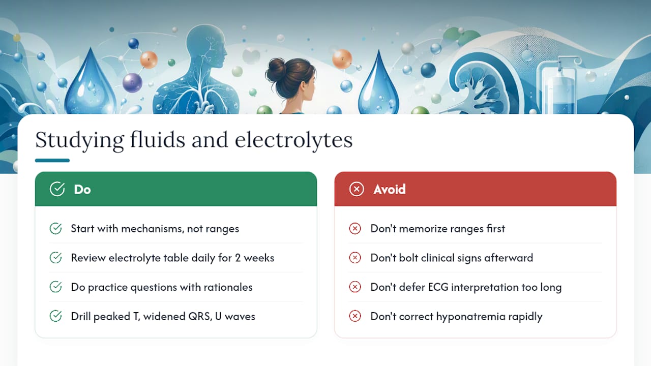
Most students approach this topic the wrong way. They memorize the normal ranges first and try to bolt clinical signs onto them afterward. That is backwards. The students who master this topic start with the mechanism: why does low potassium flatten T waves? Because potassium sets the resting membrane potential, and when it drops, the cell hyperpolarizes, making repolarization slower and the T wave smaller. Once you own that mechanism, you can reason through any potassium question you have never seen before.
The same logic applies to sodium. Hyponatremia is not just "low sodium." It is water moving into brain cells, causing them to swell. That is why the symptoms are neurologic. That is why you correct it slowly. The mechanism is the answer.
Spaced repetition works better for this topic than any other study method. The electrolyte table in Section 3 is worth reviewing daily for two weeks until the ranges and clinical pearls are automatic. Then move to worked problems: calculate a free water deficit, work through a hyperkalemia algorithm, interpret a urine sodium in the context of a clinical vignette. Practice questions with instant rationales are the fastest way to find the gaps in your reasoning, because a wrong answer with a clear explanation is more valuable than a right answer you cannot explain.
ECG interpretation is the piece most students defer too long. You do not need to read a full 12-lead to pass your exams or protect your patients. You need to recognize peaked T waves, a widened QRS, and a U wave. Those three findings, in the right clinical context, tell you everything about potassium. Drill those patterns until they are reflexive.
The Nursingschoolpro study center organizes practice questions by topic, including fluid and electrolyte imbalances, with rationales that explain the mechanism behind each answer. Progress analytics show you exactly which disorders you are missing most, so your study time goes where it matters.
Mastering fluids and electrolytes with Nursingschoolpro
Fluid and electrolyte questions appear on the NCLEX, HESI A2, and TEAS, and they show up on every clinical shift. The gap between knowing the ranges and applying them under pressure is where most students struggle. Nursingschoolpro closes that gap with practice questions mapped to every major electrolyte disorder, instant rationales that explain the mechanism (not just the answer), and real-time analytics that identify which imbalances you are consistently missing.

If you are working through this topic now, the fastest next step is a 48-hour free trial. You will get access to fluid and electrolyte practice sets, calculation drills, and the full study center. No long-term commitment. Start your free trial at Nursingschoolpro and see exactly where your knowledge gaps are before your next exam.
Sources
- Chapter 15 Fluids and Electrolytes - Nursing Fundamentals
- Fluid and Electrolyte Balance - MedlinePlus
- Hydrating for health - NIH News in Health
A note on reference ranges: Every numeric value in this article reflects commonly cited clinical standards. Always confirm against your facility's specific laboratory reference ranges, since analyzer methods and patient population norms vary.
This article is for educational purposes only and is not a substitute for clinical judgment, institutional protocols, or guidance from a licensed healthcare provider.